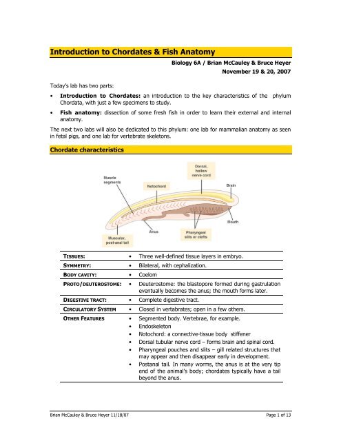
Introduction to Chordates & Fish Anatomy - De Anza College
Introduction to Chordates & Fish Anatomy - De Anza College
Introduction to Chordates & Fish Anatomy - De Anza College

You also want an ePaper? Increase the reach of your titles
YUMPU automatically turns print PDFs into web optimized ePapers that Google loves.
<strong>Introduction</strong> <strong>to</strong> <strong>Chordates</strong> & <strong>Fish</strong> Ana<strong>to</strong>my<br />
Biology 6A / Brian McCauley & Bruce Heyer<br />
November 19 & 20, 2007<br />
Today’s lab has two parts:<br />
• <strong>Introduction</strong> <strong>to</strong> <strong>Chordates</strong>: an introduction <strong>to</strong> the key characteristics of the phylum<br />
Chordata, with just a few specimens <strong>to</strong> study.<br />
• <strong>Fish</strong> ana<strong>to</strong>my: dissection of some fresh fish in order <strong>to</strong> learn their external and internal<br />
ana<strong>to</strong>my.<br />
The next two labs will also be dedicated <strong>to</strong> this phylum: one lab for mammalian ana<strong>to</strong>my as seen<br />
in fetal pigs, and one lab for vertebrate skele<strong>to</strong>ns.<br />
Chordate characteristics<br />
TISSUES: • Three well-defined tissue layers in embryo.<br />
SYMMETRY: • Bilateral, with cephalization.<br />
BODY CAVITY: • Coelom<br />
PROTO/DEUTEROSTOME: • <strong>De</strong>uteros<strong>to</strong>me: the blas<strong>to</strong>pore formed during gastrulation<br />
eventually becomes the anus; the mouth forms later.<br />
DIGESTIVE TRACT: • Complete digestive tract.<br />
CIRCULATORY SYSTEM • Closed in vertabrates; open in a few others.<br />
OTHER FEATURES • Segmented body. Vertebrae, for example.<br />
• Endoskele<strong>to</strong>n<br />
• No<strong>to</strong>chord: a connective-tissue body stiffener<br />
• Dorsal tubular nerve cord – forms brain and spinal cord.<br />
• Pharyngeal pouches and slits – gill related structures that<br />
may appear and then disappear early in development.<br />
• Postanal tail. In many worms, the anus is at the very tip<br />
end of the animal’s body; chordates typically have a tail<br />
beyond the anus.<br />
Brian McCauley & Bruce Heyer 11/18/07 Page 1 of 13
Early development of chordates<br />
The defining features of chordates appear early in development.<br />
Gastrulation<br />
Like other animals, chordates<br />
progress through blastula<br />
and gastrula stages.<br />
Gastrulation not only forms<br />
the beginning of the digestive<br />
tract, it also forms the three<br />
embryonic tissue types:<br />
endoderm, mesoderm, and<br />
ec<strong>to</strong>derm.<br />
The diagram at right shows<br />
gastrulation in a frog. The<br />
process is somewhat similar<br />
<strong>to</strong> gastrulation in echinoderms<br />
(as shown in last<br />
week’s handout on animal<br />
tissues and development).<br />
However, chordate<br />
gastrulation is somewhat<br />
more complex, partly<br />
because the early embryo is<br />
more asymmetrical and more<br />
filled in with cells.<br />
<strong>Chordates</strong> are deuteros<strong>to</strong>mes.<br />
This means that the<br />
blas<strong>to</strong>pore (the opening in<strong>to</strong><br />
the archenteron that forms<br />
during gastrulation) becomes the anus; the mouth forms later. Echinoderms are also<br />
deuteros<strong>to</strong>mes, but annelids and arthropods are pro<strong>to</strong>s<strong>to</strong>mes. In those phyla, the blas<strong>to</strong>pore<br />
becomes the mouth, and the anus forms later. This is one of the reasons that chordates are<br />
considered <strong>to</strong> be more closely related <strong>to</strong> echinoderms than <strong>to</strong> arthropods.<br />
No<strong>to</strong>chord formation<br />
The no<strong>to</strong>chord, one of the unique defining characteristics of chordates, is a semi-stiff rod of<br />
connecting tissue that forms in the embryo and <strong>to</strong> guide the development of the vertebral<br />
column (backbone) of vertebrates, along with other structures. In your own body, the no<strong>to</strong>chord<br />
has mostly disappeared; the only remnants are the cartilaginous disks between your vertebrae<br />
Neurulation<br />
Neurulation forms the dorsal hollow nerve cord in a developmental event that bears some<br />
resemblance <strong>to</strong> gastrulation. The dorsal hollow nerve cord is another unique feature of<br />
chordates.<br />
Following gastrulation, the presence of a no<strong>to</strong>chord induces neurulation of the overlying dorsal<br />
ec<strong>to</strong>derm. This third stage of morphogenesis is unique <strong>to</strong> chordates. The ec<strong>to</strong>derm above the<br />
no<strong>to</strong>chord thickens <strong>to</strong> form the neural plate. This plate then invaginates <strong>to</strong> form a furrow along<br />
the anterior-posterior axis. The folds along the groove eventually seal over the furrow <strong>to</strong> form the<br />
Brian McCauley & Bruce Heyer 11/18/07 Page 2 of 13
neural tube that in turn develops in<strong>to</strong> the dorsal hollow nerve cord. This nerve cord<br />
eventually develops in<strong>to</strong> the central nervous system, including the spinal cord and brain.<br />
By contrast, in non-chordate animals the main nerve cord is solid and usually ventral.<br />
The diagram below (fig. 47.14 from Campbell) shows neurulation and associated events in a<br />
frog.<br />
Chordate development specimens:<br />
Models of frog development & neurulation. Note the stages of development following<br />
gastrulation. Identify the subsequent appearance of the no<strong>to</strong>chord, neural plate, and<br />
finally the neural tube and neural crest. Compare <strong>to</strong> Campbell Fig. 47.14.<br />
Slides of 13-hour & 18-hour developing chick embryos. Compare the developing chick<br />
<strong>to</strong> the models of frog morphogenesis and identify the corresponding homologous structures<br />
named above. Refer <strong>to</strong> the Pho<strong>to</strong> Atlas Fig. 2.16.<br />
☛ (CAUTION: We will be examining <strong>to</strong>day several slides of this series showing stages of<br />
development in the chick embryo. The slides with the later stages are thick and can only be<br />
viewed at low power. Don’t break them by using a higher power objective.)<br />
Non-vertebrate chordates: urochordates and cephalochordates<br />
Urochordates and cephalochordates are in the phylum Chordata, but they aren’t vertebrates; in<br />
fact, you might not immediately recognize them as belonging <strong>to</strong> your own phylum.<br />
Urochordates (tunicates)<br />
Tunicates (also called sea squirts) are marine suspension feeders. They live in the ocean, pump<br />
water though their gut, and capture small particles of food suspended in the water. They are<br />
different from vertebrates in many respects:<br />
No cephalization of the dorsal nerve tube – in other words, no brain.<br />
Open circula<strong>to</strong>ry system that can even reverse its direction of flow.<br />
Brian McCauley & Bruce Heyer 11/18/07 Page 3 of 13
Pharyngeal arches and slits form a ciliated filter basket (pharynx) used for gas exchange<br />
and suspension feeding. Water is drawn in through an incurrent siphon and pumped out through<br />
an excurrent siphon, passing through the slits in the pharynx. A sheet of mucus is used <strong>to</strong><br />
capture suspended particles. The arches of the pharynx are vascularized, and serve for gas<br />
exchange as well as food capture.<br />
Many urochordates are sessile; they spend their adult lives glued <strong>to</strong> the bot<strong>to</strong>m of the ocean.<br />
The diagram above (fig.34.4 from Campbell shows adult tunicates in (a) and (b), and a larval<br />
tunicate in (c).<br />
While urochordates differ from vertebrates in many ways, they also show the defining<br />
characteristics of the phylum Chordata, including the no<strong>to</strong>chord, and the dorsal hollow nerve<br />
cord.<br />
We don’t have many urochordate specimens in the lab, but:<br />
Preserved specimens of adult ascidians. Identify the incurrent siphon, the filter<br />
basket formed by the pharyngeal arches, the atrium, and the excurrent siphon.<br />
Where is the no<strong>to</strong>chord<br />
Cephalochordates (Amphioxus, or lancelets)<br />
The lancelets (Class Cephalochordata) resemble<br />
fish larvae, but they never develop bones. They are<br />
chordates, but not vertebrates.<br />
The no<strong>to</strong>chord remains throughout life and serves<br />
as a semi-rigid endoskele<strong>to</strong>n. Mesodermal blocks<br />
develop segmented muscle bands called<br />
myo<strong>to</strong>mes. The atrial aperture (atriopore) is<br />
directed posteriorly, and the anus is located outside<br />
the atrium posterior <strong>to</strong> the atriopore. A pre-anal<br />
ventral fin and post-anal caudal fin aid mobility as<br />
the lancelet burrows tail-down in<strong>to</strong> sandy substrate<br />
projecting its oral aperture and ventral filter basket<br />
up in<strong>to</strong> the water column.<br />
The diagram at right (fig.34.5 from Campbell) shows<br />
most of the features you should be able <strong>to</strong> see in<br />
our microscope slides of a lancelet. Note that there<br />
is a pathway that water passes through, called the<br />
Brian McCauley & Bruce Heyer 11/18/07 Page 4 of 13
atrium; this is separate from the intestine. As water is pumped through the atrium, it is forced<br />
through the pharyngeal slits, where food particles are captured. The food particles are then<br />
passed in<strong>to</strong> the digestive tract.<br />
Slides with w.m. & c.s. of the lancelet Amphioxus. Identify the following structures:<br />
tentacles, mouth, no<strong>to</strong>chord, dorsal nerve cord, pharynx with slits, atrium, intestine, tail,<br />
muscle segments (myo<strong>to</strong>mes).<br />
Vertebrates<br />
The vertebrates (phylum Chordata, subphylum Vertebrata) include fish, amphibians, reptiles<br />
(including birds) and mammals. All these animals have the basic characteristics of chordates,<br />
with some added twists:<br />
• The anterior region of the dorsal hollow nerve cord expands <strong>to</strong> become a brain and is<br />
encased within a skeletal cranium.<br />
• Mesodermal blocks develop not only myo<strong>to</strong>mes, but a segmented vertebral column that<br />
protects the posterior region of the dorsal nerve cord (spinal cord) and replaces the<br />
no<strong>to</strong>chord. The cranium and vertebral column comprise the axial skele<strong>to</strong>n of vertebrates<br />
allowing an elaborate central nervous system and powerful muscle action with a flexible<br />
body.<br />
• The development of a brain has further spurred the development of more sophisticated<br />
cephalic sensory organs, especially eyes, nostrils and ears.<br />
• To support their enhanced homeostasis, vertebrates also have a closed circula<strong>to</strong>ry system<br />
with distinctive types of heart and kidneys, and a greater suite of endocrine organs.<br />
Vertebrate specimens:<br />
Slides of 21-hour through 48-hour developing chick embryos. Observe the dramatic<br />
cephalization of the dorsal hollow nerve cord with the distinction of brain from spinal<br />
cord and development of cephalic sensory organs, notably the eyes and ears. Note also the<br />
segmentation evident in the developing myomeres, and the early establishment of the<br />
segmented vertebral column.<br />
Slides of 2-day through 7-day developing chick embryos. Observe the limb buds<br />
developing in<strong>to</strong> the appendicular skele<strong>to</strong>n. Locate each of the four extra-embryonic<br />
membranes and describe their respective functions. C.f., Campbell Fig. 47.17.<br />
Slide of lamprey ammocoetes larva. The ammocoete larva of lampreys resembles a<br />
lancelet in form and habit, but with the vertebrate modifications described above.How is this<br />
larva similar <strong>to</strong> the lancelet How is it different How does it differ from the adult lamprey<br />
Skele<strong>to</strong>n of a bony fish. Note the segmented axial endoskele<strong>to</strong>n. What is the function for<br />
the spines protruding from the vertebral segments Examine the hinged jaw, buccal<br />
chamber, and pharynx with gill arches. List and locate the unpaired and paired fins.<br />
Skele<strong>to</strong>n of a shark. Note the corresponding structures <strong>to</strong> those listed for the boney fish.<br />
How is the shark skele<strong>to</strong>n different from the boney fish (composition; attachment of fins)<br />
In a later lab, you’ll look at vertebrate skele<strong>to</strong>ns in more detail, contrasting fish with mammals<br />
and other vertebrates.<br />
Brian McCauley & Bruce Heyer 11/18/07 Page 5 of 13
<strong>Fish</strong> Ana<strong>to</strong>my<br />
This part of the lab comes <strong>to</strong> you straight from the grocery s<strong>to</strong>re. You’ll have the opportunity <strong>to</strong><br />
examine a variety of fresh fish.<br />
<strong>Fish</strong> Taxonomy<br />
The taxonomy of <strong>Chordates</strong> can be confusing, because there are traditional, well-known groups<br />
(like fish) that don’t reflect real evolutionary relationships. Most fish have traditionally been<br />
grouped in two classes within the phylum Chordata: Osteichtyes, or bony fish (which includes<br />
most kinds of fish) and the Chondrichthyes, or cartilaginous fish (which includes sharks and<br />
rays). However, the actual evolutionary relationships of fish are a bit more complex, as shown in<br />
the cladograms in Campbell, ch. 34.<br />
This lab will focus on the bony fish, traditionally classified as Osteichthyes. Most of the bony fish<br />
belong <strong>to</strong> a group within the Osteichthyes called the Actinopterygii.<br />
<strong>Fish</strong> Ana<strong>to</strong>my<br />
In this handout, you’ll see various diagrams with numerous ana<strong>to</strong>mical features listed. Study<br />
them all, but the terms you’re likely <strong>to</strong> see on a lab exam are those listed in bold type in the text<br />
of this handout. You won’t be tested on everything in the diagrams.<br />
External ana<strong>to</strong>my<br />
Before you cut the fish open, see what you can learn from the outside. Note that different fish<br />
may have very different shapes, especially for their fins. Can you make any guesses about how<br />
your fish lives<br />
Look for the features shown in the diagram below.<br />
Brian McCauley & Bruce Heyer 11/18/07 Page 6 of 13
Fins: Note the caudal, dorsal, pelvic, pec<strong>to</strong>ral, and anal fins. You’ll see very different shapes of<br />
these fins in the various fish we have in lab.<br />
Lateral line: You may see a pigmented stripe marking the location of the lateral line sense<br />
organs. Can you see any evidence of the lateral line canals or organs<br />
Inside the mouth: Look inside the mouth. Note where the water would go as it passes over the<br />
gills, and note where the food would go when the fish eats. Take a look at the <strong>to</strong>ngue. How is it<br />
different from your <strong>to</strong>ngue Note the teeth. Compare the teeth of the various fish in the room.<br />
<strong>Fish</strong> teeth come in a variety of syles, some of which are shown in the diagram on the next page.<br />
In addition <strong>to</strong> the teeth in the jaws, fish often have pharyngeal teeth located back in the<br />
throat. The pharyngeal teeth are derived from pharyngeal arches, one of the key chordate<br />
characteristics. Why do you suppose they would have these “extra” teeth<br />
While you’re looking in the mouth, look at the gill arches and the operculum. The gill arches<br />
are pharyngeal arches that support and protect the gills. The gill arches usually have gill rakers<br />
on the anterior (front) side. Gill rakers are small projections sticking out of the gill arch; they<br />
prevent the fish’s food from escaping out through the gills. <strong>Fish</strong> that eat large prey have widely<br />
spaced gill rakers; fish that eat tiny zooplank<strong>to</strong>n have closely spaced, filter-like rakers.<br />
Brian McCauley & Bruce Heyer 11/18/07 Page 7 of 13
I have no idea what the captions say on the above figure (from<br />
http://gyeonggi.go.kr/fishs/fishs2.php). However, it’s a picture of a gill arch with gills on the<br />
posterior side (the right in this picture) and gill rakers on the anterior side (left).<br />
Note how opercular pumping might work in your fish. What would make the water enter the<br />
mouth What would make it pass out across the gills<br />
Nostrils: where does the water go when it passes through the fish’s nostrils<br />
Vent: <strong>Fish</strong> have a single opening, the vent, that serves as the anus and the urogenital opening.<br />
(Urogenital means urinary and genital – that is, the opening through which eggs or sperm pass<br />
during reproduction). Find the vent in your fish. The vent marks the posterior end of the body<br />
cavity; behind that, the fish is all tail.<br />
Gills<br />
Recall the structure of fish gills:<br />
Cut away the operculum so you have a good look at the gills and the inside of the mouth. Each<br />
gill is supported by a bony gill arch, with the spiny gill rakers projecting forward. Note the<br />
filaments of the gills, each with many lamellae. You will be able <strong>to</strong> see this better under the<br />
dissecting microscope.<br />
The fish’s blood must flow through the gills <strong>to</strong> get oxygenated, so the gills are filled with blood<br />
vessels. The heart is located just behind the gills, but you won’t be able <strong>to</strong> see it until you cut the<br />
body open. You’ll come back <strong>to</strong> the heart in a little while.<br />
Inside the body cavity<br />
Cut open the fish’s body along the ventral (belly) side. You’ll see all the internal organs sitting in<br />
the coelom. To get a better view, cut away one side of the body wall, removing the muscle,<br />
bone, and skin so you have an unobstructed view. Note the organs shown in the diagram below<br />
(diagram is from Campbell, ch. 34.)<br />
Brian McCauley & Bruce Heyer 11/18/07 Page 8 of 13
Digestive tract. The digestive tract of fish is similar <strong>to</strong> that of mammals. Food passes from the<br />
mouth down the esophagus <strong>to</strong> the s<strong>to</strong>mach. You may be able <strong>to</strong> see pyloric ceca, which<br />
function as accessory digestive glands (mammals don’t have these). The liver is large; it<br />
secretes bile in<strong>to</strong> the gall bladder, from which the bile passes in<strong>to</strong> the intestine. The intestine<br />
is fairly long. You may find remnants of food in the s<strong>to</strong>mach or the intestine.<br />
Just like mammals, fish have digestive tracts that are adapted <strong>to</strong> the food they eat. Herbivorous<br />
fish generally have longer intestines <strong>to</strong> aid in digestion and nutrient absorption from their low<br />
calorie food. Some also have a gizzard, a muscular organ that helps <strong>to</strong> mash up the food for<br />
better digestion.<br />
Brian McCauley & Bruce Heyer 11/18/07 Page 9 of 13
Swim bladder. The swim bladder is a large light-colored structure near the dorsal side of the<br />
body cavity. Bony fish can adjust their buoyancy by putting gas in<strong>to</strong> the swim bladder. The most<br />
abundant gas in the swim bladder is oxygen, which comes from the blood. A specialized gas<br />
gland generates lactic acid, acidifying the blood in the gas gland and causing it <strong>to</strong> release its<br />
oxygen (remember the Bohr shift). This gland is accompanied by a specialized countercurrent <strong>to</strong><br />
maintain the incredibly strong gradient of oxygen partial pressure between the swim bladder and<br />
the blood. Some fish can have over 100 atmospheres of oxygen partial pressure in the swim<br />
bladder, but their blood can never contain more O 2 pressure than the surrounding water, which<br />
never contains more O 2 pressure than the air (0.2 atm).<br />
Kidney & urinary bladder. Does your fish live in salt water or in fresh water Either way it has<br />
a kidney, but the function of the kidney is quite different in these two environments. The kidney<br />
is long and thin and dark; it is located along the very <strong>to</strong>p of the body cavity. Urine produced by<br />
the kidney enters the urinary bladder, which exits in<strong>to</strong> the same area as the anus and the gonad.<br />
Gonad. Most fish are either male or female, though some change sexes. You should be able <strong>to</strong><br />
see either an ovary, which may be filled with large orange eggs, or a testis, filled with white<br />
sperm.<br />
Circula<strong>to</strong>ry system. Remember that fish pump their blood once per circuit. The heart is<br />
located close <strong>to</strong> the gills, and blood passes through the gills and then through the systemic<br />
circulation before making it back <strong>to</strong> the heart. You may have <strong>to</strong> do a little extra dissection <strong>to</strong> find<br />
the heart; it’s typically just behind and beneath the gills. Note the direct blood flow from heart <strong>to</strong><br />
gills.<br />
Other ana<strong>to</strong>mical features<br />
Muscle. <strong>Fish</strong> have a lot of muscle. If you peel away the skin, you can see the sections of muscle<br />
that are familiar <strong>to</strong> anyone who eats fish. The muscle of fish provides a very clear example of the<br />
segmented mesoderm that is characteristic of chordates. Also note the rays of the fins. Most<br />
fish can pull their fins flat <strong>to</strong> the body or extend them. The fins are extended by rays that<br />
penetrate down in<strong>to</strong> the fish’s muscle; the flexing muscle levers the rays in<strong>to</strong> an upright position.<br />
Eyes. If you’re good at dissecting small things, you may be able <strong>to</strong> sort out the ana<strong>to</strong>my of the<br />
eye. It’s a bit squishy, but you may be able <strong>to</strong> find some of the features shown below:<br />
Brian McCauley & Bruce Heyer 11/18/07 Page 10 of 13
Note that in fish eyes there is usually a tapetum behind the retina; this silvery reflective layer<br />
bounces light back <strong>to</strong> the pho<strong>to</strong>recep<strong>to</strong>r cells in the retina, allowing the fish <strong>to</strong> see better in low<br />
light. Many mammals also have this feature, but humans don’t.<br />
Brain. It takes a little work with scissors, but you can cut open the brain case and find the brain<br />
inside. You’ll probably have a hard time locating the different regions of the brain, but here’s a<br />
diagram:<br />
Note that there are large semicircular canals (balance organs) associated with the brain.<br />
Skele<strong>to</strong>n. <strong>Fish</strong> live in the water, and they don’t have <strong>to</strong> support all their weight with their<br />
skele<strong>to</strong>ns. For this reason, fish skele<strong>to</strong>ns are generally much weaker than mammalian skele<strong>to</strong>ns.<br />
That’s why you can cut the skull open with scissors. Compare your fish <strong>to</strong> the mounted fish<br />
skele<strong>to</strong>ns in lab. We’ll look at skele<strong>to</strong>ns in more detail in a later lab.<br />
Brian McCauley & Bruce Heyer 11/18/07 Page 11 of 13
Brian McCauley & Bruce Heyer 11/18/07 Page 12 of 13
Terms and structures <strong>to</strong> remember:<br />
There are two parts <strong>to</strong> this lab: chordates in general, and the ana<strong>to</strong>my of fish.<br />
<strong>Chordates</strong><br />
• Appendicular skele<strong>to</strong>n<br />
• Atrium<br />
• Axial skele<strong>to</strong>n<br />
• Cephalochordate<br />
• Chordata<br />
• Cranium<br />
• <strong>De</strong>uteros<strong>to</strong>me<br />
• Dorsal hollow nerve cord<br />
• Gastrulation<br />
• Myo<strong>to</strong>mes (muscle segments)<br />
• Neurulation<br />
• No<strong>to</strong>chord<br />
• Pharynx; pharyngeal slits; pharyngeal basket<br />
• Tail<br />
• Urochordates<br />
• Vertebral column<br />
• Vertebrate<br />
<strong>Fish</strong> Ana<strong>to</strong>my<br />
You should be able <strong>to</strong> identify the following structures in any of the fish that are in <strong>to</strong>day’s lab<br />
(remember, other groups may have different species of fish – you should look at them all).<br />
• Fins: pec<strong>to</strong>ral, pelvic, dorsal, anal, caudal<br />
• Gills, gill arches, gill rakers<br />
• Gonad (ovary or testis)<br />
• Heart<br />
• Intestine<br />
• Kidney<br />
• Lateral line<br />
• Liver & gall bladder<br />
• Operculum<br />
• Pyloric ceca<br />
• S<strong>to</strong>mach<br />
• Swim bladder<br />
• Teeth and pharyngeal teeth<br />
• Vent<br />
Brian McCauley & Bruce Heyer 11/18/07 Page 13 of 13