Download

1 / 38
390 likes | 408 Views
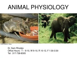
ANIMAL PHYSIOLOGY. BIOL 3151: Principles of Animal Physiology. Dr. Tyler Evans Email: tyler.evans@csueastbay.edu Phone: 510-885-3475 Office Hours: M,W 10:30-12:00 or appointment Website: http ://evanslabcsueb.weebly.com /. LAST LECTURE. Why do squid have giant axons?.

E N D
ANIMAL PHYSIOLOGY BIOL 3151: Principles of Animal Physiology Dr. Tyler Evans Email: tyler.evans@csueastbay.edu Phone: 510-885-3475 Office Hours: M,W 10:30-12:00 or appointment Website: http://evanslabcsueb.weebly.com/
LAST LECTURE Why do squid have giant axons? • axons that activate muscles at the far end of the mantle have VERY LARGE DIAMETERS • axons that activate muscles in the region of the mantle closest to the central nervous system have smaller diameters • combining axons of various diameters allows the near-simultaneous contraction of the mantle
LAST LECTURE TRADE-OFFS OF GIANT AXONS? • although increasing axon diameter provides increases in conduction velocity, there are two main disadvantages to using large axons to increase conduction velocity: • Large axons take up more space and this may limit the number of neurons that can be packed into the nervous system • Large diameter axons have a much larger volume of cytoplasm per unit length, making them energetically expensive to make and maintain.
LAST LECTURE MYELIN SHEATHS • true myelin sheaths were an important evolutionary innovation • allowed rapid signal conduction in a small amount of space • helped to provide conditions for complex vertebrate nervous systems • more complex nervous systems allowed animals to evolve more complex behavior, physiology, social systems, etc. Mammals (lots of myelin) Lampreys/Hagfish (no myelin)
LAST LECTURE ELECTRICAL VS CHEMICAL SYNPASES • ELECTRICAL SYNAPSES and CHEMICAL SYNAPSES differ in a number of ways: • DIRECTION OF FLOW OF INFORMATION • in electrical synapses, information can flow in BOTH DIRECTIONS • because pre- and post-synaptic cells are directly connected • SPEEED OF TRANSMISSION • electrical synapses are fast • chemical synapses are slower because of delays associated with the diffusion of neurotransmitters across the synaptic cleft and their binding to receptors on the post-synaptic neuron
TWO EXCELLENT STUDENT QUESTIONS FROM NEUROPHYSIOLOGY LECTURES Does the thickness of myelin differ between axons? YES! • vertebrates have axons that vary in diameter, though not to the same degree as we see in squid • large diameter axons have thick myelin sheaths and small diameter axons have thin myelin sheaths • ratio between axon diameter and myelin thickness is constant (0.6-0.7) http://www.nature.com/scitable/topicpage/myelin-a-specialized-membrane-for-cell-communication-14367205
TWO EXCELLENT STUDENT QUESTIONS FROM NEUROPHYSIOLOGY LECTURES • 2. Which type of synapse is more energetically demanding: chemical or electrical? • chemical synapse require more energy due to formation and transport of SYNAPTIC VESICLES • vesicles can be recycled, but still more energy demanding http://www.albany.edu/faculty/cafrye/apsy601/Ch.04feb10,psychopharmacology.html
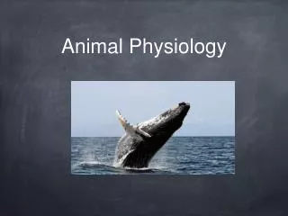
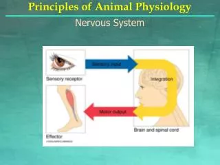
TODAY’S LECTURE CELLULAR MOVEMENT AND MUSCLES • muscles are a distinctive cell type found only in animals • simplest animals lack true muscles, but do have cells that contract • what’s full of holes but can hold water?
CELLULAR MOVEMENT AND MUSCLES • muscle-like cells first arose in Cnidarians such as Hydra • the Hydra uses MYOEPITHELIAL CELLS to support and extend their body stalk
CELLULAR MOVEMENT AND MUSCLES • true muscles first appeared in a group of animals call ctenophores (comb jellies) • ctenophores like sea walnuts (right) and sea goose berries have true smooth muscles in their bodies walls that they use for locomotion
CELLULAR MOVEMENT AND MUSCLES • although these primitive animals have several different types of muscles, complex animals display much greater diversity in muscle anatomy and physiology • the greatest muscle diversity occurs in the vertebrates • with the transition to land and the challenges of movement under the full weight of gravity, muscle diversified rapidly and became specialized for many different processes Tiktaalik fossil represents an important group of vertebrates involved in the transition toward life on land
CELLULAR MOVEMENT AND MUSCLES • regardless of the type of movement, the same intracellular machinery underlies each one • a quote from your textbook: “when you marvel at the athleticism of a cheetah sprinting, a tuna swimming or a hummingbird hovering, remember these impressive capacities depend on cellular machinery not unlike that found in the fungus growing on your shower curtain”
CELLULAR MOVEMENT AND MUSCLES • common machinery is the CYTOSKELETON and associated MOTORPROTEINS • microtubules and microfilaments are most important of these components • diversity in cellular movement is possible because these basic elements can be arranged and used in countless combinations • three general ways cells use these elements to move: 1. use cytoskeleton like a “roadway” (most common) • motor proteins act as trucks carrying cargo over cytoskeleton roadways • e.g. transport of proteins between different organelles cargo motor protein cytoskeleton Textbook Fig 5.1 pg 198
CELLULAR MOVEMENT AND MUSCLES • three general ways cells use these elements to move: 2. Cytoskeletal fibers act like a “bulldozer” to push cellular contents forward • often referred to as AMOEBOID movement and is common in protists • e.g. cells involved in the immune response use amoeboid movement motor protein cargo cytoskeleton Textbook Fig 5.1 pg 198
CELLULAR MOVEMENT AND MUSCLES • three general ways cells use these elements to move: 3. Motor proteins pull a cytoskeleton rope • cells then reorganize in cytoskeleton so that this pulling translates into movement • e.g. these types of cytoskeletal structures are the basis of cilia, flagella and muscle motor protein cargo cytoskeleton Textbook Fig 5.1 pg 198
CELLULAR MOVEMENT AND MUSCLES • MICROTUBULESand MICROFILAMENTSare most important of components of cell movements MICROTUBULES • microtubules are so named because of their tube-like appearance and are composed of long strings of the protein TUBULIN, which is a dimer of two closely related proteins alpha (α) tubulin and beta (β) tubulin • the first step of tubulin assembly occurs when αand β-tubulin combine • GTP (stored energy) is bound to α tubulin • α and β-tubulin combine with a reaction that uses the stored energy of GTP • result isα-tubulin now has a negative charge relative to β-tubulin • charge difference is key to assembly Textbook Fig 5.4 pg 201
CELLULAR MOVEMENT AND MUSCLES MICROTUBULES • like magnets, the positive and negative ends of tubulin attach to form a growing chain or PROROFILAMENT • protofilaments then line up to form a sheet that eventually rolls into a tube to form a complete MICROTUBULE Textbook Fig 5.4 pg 201
CELLULAR MOVEMENT AND MUSCLES MICROTUBULES • most cells arrange their microtubules like spokes on a wheel, radiating out to the periphery of the cell from a central MICROTUBULE ORGANIZING CENTER • cells use this microtubule network to control the movement of vesicles and other cargo to different parts of the cell “-” end of the microtubule is closest to the microtubule organizing center, while the “+” end is closest to the cell periphery Textbook Fig 5.6 pg 202
CELLULAR MOVEMENT AND MUSCLES MICROTUBULES • cells use this microtubule network to control the movement of vesicles and and other cargo to different parts of the cell • e.g. color change in camouflaged animals • Xenopus frog darkens its skin by transporting pigment granules from the MICROTUBULE ORGANIZING CENTER to the periphery of the skin along microtubules Textbook Fig 5.3 pg 200
CELLULAR MOVEMENT AND MUSCLES MICROTUBULES • microtubules are involved in important cell movements required for survival • things like cell division, axon structure, movement of cilia and flagella Textbook Table 5.1 pg 206
CELLULAR MOVEMENT AND MUSCLES MICROTUBULES • microtubules are very sensitive to temperature: they will disassemble if too cold • not a problem for ENDOTHERMS (warm-blooded animals), but ECTOTHERMS (cold-blooded animals) can live in very cold environments (e.g. polar regions) • microtubules assemble at lower temperatures in ectotherms from cold environments • requires only single mutation in tubulin gene to promote cold stability • animals in living in the Antarctic Ocean have adaptations that prevent microtubules from disassembling in the cold water (-2°C)
CELLULAR MOVEMENT AND MUSCLES TRANSPORT USING MICROTUBULES • if microtubules are like roads, how does cargo know which direction to travel? • orientation of α and β-tubulin give tubulin POLARITY (a charge difference) • the negative ends stay near microtubule organizing center and the positive ends stretch towards the cell periphery • MOTOR PROTEINS (proteins that move along microtubules) recognize this polarity and each motor protein moves in a characteristic direction Textbook Fig 5.4 pg 201
CELLULAR MOVEMENT AND MUSCLES TRANSPORT USING MICROTUBULES • motor proteins recognize this polarity and each motor protein moves in a characteristic direction: • KINESIN: moves along the microtubule in the POSITIVE direction • DYNEIN: moves along the microtubule in the NEGATIVE direction KINESIN DYNEIN
CELLULAR MOVEMENT AND MUSCLES TRANSPORT USING MICROTUBULES e.g. neurotransmitters transport down axons • neurotransmitters are released at synapses to induce a response • neurotransmitters are carried from the cell body (i.e. soma) down the axon on microtubules • kinesin can transport neurotransmitters to the end of the synapse (+ direction) • dynein then carries the empty synaptic vesicle back to the soma (- direction) textbook Fig 4.16 pg 162 textbook Fig 5.7 pg 204
CELLULAR MOVEMENT AND MUSCLES TRANSPORT USING MICROTUBULES e.g. neurotransmitters transport down axons textbook Fig 5.7 pg 204
CELLULAR MOVEMENT AND MUSCLES MICROTUBULES • CILIA and FLAGELLA rely on microtubules to generate movement • microtubules in cilia and flagella are arranged into a structure called the AXONEME • each doublet microtubule around the periphery of the axoneme contains a dynein motor-a signal triggers dynein to stretch out and attach to negative end of neighboring tubulin, then pulls itself forward • by altering this movement between opposite sides of the axoneme, a whip-like movement is generated textbook Fig 5.8 pg 205
CELLULAR MOVEMENT AND MUSCLES MICROFILAMENTS • microfilaments are the other type of cytoskeletal elementused in movement • also involved in transport within cells, but in cell shape changes and moving from place to place • microfilament based movement uses ACTINand the motor protein MYOSIN • microfilaments are composed of long string of ACTIN • microfilaments form in much the same way microtubules: “+” and “-” ends of actin assemble • actin monomers are termed G-ACTIN • “G” stands for globular • referred to as F-ACTIN when in polymers • “F” stands for filamentous
CELLULAR MOVEMENT AND MUSCLES MICROFILAMENTS • actin polymerization (formation of F-actin) can generate cell movement • important in two types of amoeboid movement in cells: • 1. FILIPODIA: thin extensions of cells • digestive cells use filipodia to build microvilli • cells of the small intestine use microvilli to increase surface areas of cells responsible for absorbing nutrients from food
CELLULAR MOVEMENT AND MUSCLES MICROFILAMENTS • actin polymerization (formation of F-actin) can generate movement • important in two types of amoeboid movement in cells: Lamellipodium • 2. LAMELLIPODIA:are sheet-like protrusion of the cell and are involved in cell migration, for example during development
CELLULAR MOVEMENT AND MUSCLES MICROFILAMENTS • sperm also use actin polymerization during fertilization • fertilization depends on sperm controlling growth of actin toward the egg • sperm use actin polymerization to push an extension of their plasma membrane into the egg • once this occurs the sperm can transfer nuclear DNA to the egg • Textbook Fig 5.11 pg 208
CELLULAR MOVEMENT AND MUSCLES MICROFILAMENTS • although possible to use actin polymerization for movement, in most situations microfilaments are used in combination with motor protein MYOSIN • there are many ISOFORMS of myosin (17 actually), but the structure is similar in diverse organisms: head, tail, neck HEAD: possesses ATPase activity, which provides the energy required for movement NECK: allows regulatory proteins to bind to myosin and modify its activity TAIL: binds cargo, like vesicles, organelles, or membranes • Textbook Fig 5.12 pg 209
CELLULAR MOVEMENT AND MUSCLES MICROFILAMENTS • MOTOR PROTEINS like MYOSIN (but also KINESIN and DYNEIN) use ATPase activity to convert the energy stored in ATP into mechanical energy. • the series of events that culminate in actin-myosin based movement is called the SLIDING FILAMENT MODEL • this model can be used to describe many different types of movement involving myosin from vesicular transport to muscle contraction A useful analogy for actin-myosin based movement
CELLULAR MOVEMENT AND MUSCLES MICROFILAMENTS WHAT DOES THE ROPE REPRESENT? WHAT DOES YOUR ARMS REPRESENT? WHAT DO YOUR HANDS REPRESENT? WHICH PART REQUIRES MOST ATP?
CELLULAR MOVEMENT AND MUSCLES SLIDING FILAMENT MODEL • the myosin molecule extends by straightening its NECK (i.e. arms extending) • the myosin HEAD then forms a bond with the actin filament (i.e. hands grasping onto the rope) • this strong interaction between actin and myosin is called a CROSS BRIDGE • myosin bends and pulls the actin filament towards its tail (i.e. pull up) • this step is called the POWER STROKE • HEAD then uncouples from actin and myosin returns to the resting unattached position Actual movement depends on whether it is actin or myosin that is free to move • Textbook Fig 5.12 pg 209 • if rope attached, you will pull yourself (myosin is mobile) • If rope not attached, you will pull the rope (actin is mobile)
CELLULAR MOVEMENT AND MUSCLES • 1. UNITARY DISPLACEMENT-corresponds to the distance myosin steps during each cross bridge cycle • in rope analogy, depends on length of arms • for myosin depends of length of the NECK • myosin step are variable, but on average cover 36 nm per step Unitary displacement • Textbook Fig 5.12 pg 209
CELLULAR MOVEMENT AND MUSCLES SLIDING FILAMENT MODEL • ATP provides the chemical energy for the movement of actin and myosin • the hydrolysis of ATP causes the neck of myosin to extend forward and grasp further up the actin filament • energy released from this hydrolysis allows myosin to pull actin filament • the head remains attached until another ATP binds, which causes neck to extend once again. • if no ATP present remains myosin head remains attached to actin. This is the cause of RIGOR MORTIS • animal dies, ATP is depleted and muscle remain locked
LECTURE SUMMARY • muscles are a distinctive cell type found only in animals: true muscles first appeared in a group of animals call CTENOPHORES (comb jellies). • regardless of the type of movement, the same intracellular machinery underlies each one • microtubules and microfilaments are most important components of cellular movement • orientation of α and β-tubulin give microtubules POLARITY(a charge difference) and microtubule motor proteins (kinesin, dynein) recognize this polarity and each motor protein moves in a characteristic direction • foundation of microfilament based movement is ACTIN and the motor protein MYOSIN • the series of events that culminate in actin-myosin based movement is called the SLIDING FILAMENT MODEL • UNITARY DISPLACEMENT-corresponds to the distance myosin steps during each cross bridge cycle
NEXT LECTURE Muscle Structure and Regulation of Contraction