Spotting spots …

A recurring theme of this blog has been that the process of “seeing” is one that requires a mind that is preconditioned to process the signals passed to it from the optic nerve.  For much of the natural history that we see on a country walk, that link is already in place: we notice trees, birds, butterflies and so on, even if we can’t always put a name on what we are looking at.   That means that natural history becomes a closed loop: noticing birds means we are more likely to notice differences amongst birds – whether in type or habit or the timing of their visits – which, in turn, stimulates greater mental awareness.   But not noticing something is equally self-perpetuating and many of my posts have focussed on the natural history that most of us routinely overlook.

A good reason for not noticing aspects of natural history is that the organisms in question do not even look “alive” in the first place.  Another is that they are so tiny that they are either microscopic or only just apparent with the naked eye.   As this post deals with a group of algae that fit both of these criteria, you can be forgiven for not knowing about these organisms before, but also for wondering what kind of person scratches away at submerged stones in the counterintuitive hope of finding life.

Earlier posts have talked about algae that form crusts on rocks, such as the red algae Hildenbrandia rivularis(see “More about red algae”) or the cyanobacteria of the genus Chamaesiphon (see “A bigger splash …”).  Chamaesiphon tends to form brown to almost black spots on rocks but there is one other type of alga that also forms dark brown patches of rock in streams that I have not previously written about.  This is the brown alga Heribaudiella fluviatilis.    Most of the brown algae are marine and largely beyond the scope of this blog (but see “Swimming in a sea of ignorance”) and there are just two genera recorded from UK freshwaters.   Both are rarely recorded, but probably more common than we think.  I’ll come back to this later in the post, but freshwater brown algae really do encapsulate the conundrum in the first paragraph: not noticing becomes self-perpetuating as we repeatedly fail to realise that there is something we ought to notice.

The photos at the top of the post – taken by Wolfgang Schülz in Germany – show characteristic colonies of Heribaudiella fluviatilis, with distinct margins, in contrast to Chamaesiphon crusts, whose margins are less clearly delimited.  The best way to confirm that you are looking at Heribaudiella rather than Chamaesiphon, however, is to scrape up the crust and examine it under a microsope.  Then the characteristic cells arranged in short filaments, each with several yellow-brown chloroplasts, should be obvious.   

A microscopic view of filaments of Heribaudiella fluviatilis.  Photo by Chris Carter.  The pictures at the top of the post show macroscopic views of Heribaudiella on stones from a river in Germany (courtesy of Wolfgang Schülz).

The late Nigel Holmes was good at spotting Heribaudiella colonies (which can be just a couple of millimetres across) on stones in rivers but the ability (or, perhaps, the inclination) to notice these has declined thereafter.   We know it is out there because the primers we use for molecular studies of diatoms also appear to pick up Heribaudiella as by-catch, and we were surprised at how often it appeared.   Curiously, these primers also differentiated between Heribaudiella and another brown algal genus, Bodenella.  I wrote about Bodenella in Depths of Imagination and also mentioned that there was some dispute about whether this was truly distinct from Heribaudiella.   Last summer, I revisited a stream from which this “Bodenella” was recorded but failed to get enough material to make a satisfactory examination of its microscopic features.   The jury is out on this, especially as the habitats where we’ve detected “Bodenella” are so different to the locations where it has been securely identified elsewhere in Europe.   Personally, I would be reluctant to add this as a new UK record without having backed up a molecular identification with some more traditional taxonomy.  

Having noticed that we’re not noticing Heribaudiella fluviatilis, one of the best arguments for stimulating interest in this organism is that it tells us useful information about the state of the stream in which it is growing.  Most of the chemical data describing its habitat that I’ve seen would suggest that it prefers “good status” water and is, thus, a sign that the ecosystem is healthy.   In the final analysis, though, records of Heribaudiellaprobably say more about the quality of the observer than of the habitat.   

References

Koletić, N., Alegro, A., Vuković, Rimac, A. & Šegota, V. (2018).  Spotting the spots: the freshwater brown algaHeribaudiella fluviatilis (Areschoug) Svedelius within stream communities of southeastern Europe.   Cryptogamie, Algologie 39: 449-463.

(this paper, interestingly, notes an association between Heribaudiella and Hildenbrandia that is also discussed in Depths of Imagination)

Wehr, J.D. (2011).  Phylum Phaeophyta.  pp. 354-357.   In: The Freshwater Algal Flora of the British Isles(edited by D.M. John, B.A. Whitton & A.J. Brook).  Cambridge University Press, Cambridge. 

Wehr, J.D. & Stein, J.R. (1985).  Studies on the biogeography and ecology of the freshwater phaeophycean alga Heribaudiella fluviatilis.  Journal of Phycology 21: 81-93.

The work that we described in Depths of Imagination was recently published:

Schülz, W., Kelly, M.G., King, L. & Cantonati (2021).  Did Zebra mussel fill the type habitat of a worldwide-rare freshwater brown macroalga?  Aquatic Conservation: Marine and Freshwater Ecosystems 31: 3657-3659.

Wrote this whilst listening to: Hildegaard of Bingen, and Nick Cave’s Murder Ballards.

Currently reading: Pen Vogler’s Scoff, about the history of British food.

Cultural highlight: Munich: Edge of War – film based on Robert Harris’ book set around the Munich crisis of 1938 and starring George Mackay and Jeremy Irons.  

Culinary highlight: mutton steak from our local organic butcher, with homemade chips and peppercorn sauce.

Depths of imagination …

I’ve written many posts over the years about algae that grow in lakes, mostly based on samples I’ve collected whilst paddling around in the shallow littoral zone.   One feature that I have not previously mentioned is that the attached algal flora changes quite dramatically as you descend into the deeper parts of the lake.  These habitats have only been thoroughly explored since SCUBA diving became possible in the middle of the twentieth century.   Before then, what was known of underwater algae came from chunks of rock dredged up from depths in nets.  

This was the case in Lake Constance, which straddles the borders of Germany, Austria and Switzerland and, in particular, in the northern arm, the Überlinger See, where there are steep underwater cliffs composed of Molasse (a sedimentary sandstone/shale association).   Studies by Lauterborn and Zimmerman revealed crusts and mats smothering these cliffs and extending to great depths.   As you descended from the shallower, lighter water to the depths the composition shifted from predominately green algae to brown and red algae.  Zimmerman captured these changes in a sketch of the algae on a ledge on the cliff wall at about fifteen metres depth, showing the red alga Hildenbrandia rivularis tucked away in the shady recesses whilst Cladophora profunda (now regarded as a synonym of Aegagropila linnaei, which we met in “A load of balls …”) was more abundant on the lighter, more exposed faces.   The big interest, though, were the extensive growths of a freshwater brown alga, Bodanella lauterbornii.  

A reconstruction of the algal communities at approximately 15-20metres depth in Lake Constance at the time of Zimmermann’s survey in 1925.   a: Aegagropia linnaei; b: Bodanella lauterbornii; c: Hildenbrandia rivularis.   The foreground of the picture frame represents about 30 centimetres.   The picture at the top shows the surface of Lake Constance at the time of the modern survey (photo: Lydia King).   

The Phaeophyta – brown algae – are much more common in marine environments than in freshwaters.   A few species can be found but these form small, inconspicuous growths that are frequently overlooked.    Lauterborn and Zimmerman’s record of a rare brown algae in the depths of Lake Constance (it has only been recorded in three other lakes in Austria) came to light during the preparation of a Red List of algae in Baden-Württemberg and led to a search to see if it was still present.  

The study proved fruitless, with all the submerged cliffs covered not by algae but by extensive growths of invasive mussels, mostly zebra mussels, Dreissena polymorpha.  My illustration tries to convey the extent to which zebra mussels smother the surfaces but is still an underestimate, compared to the photographs taken by divers in this study, which show mussels filling all available spaces and often growing over each other.  Zebra mussels were first recorded in Lake Constance in the 1960s and it is possible that the algal assemblages described by Zimmerman were already declining at this point.   In the period since the original surveys, Lake Constance received considerable nutrient enrichment which would have boosted phytoplankton growth, in turn reducing the limited light available to the deep water algae yet further.  So we can envisage a two-stage decline: first gradual darkening of an already very shaded habitat followed by a coup de grace of mussels covering all available surfaces.   

An impression of Bodenella lauterbornii, based on illustrations in Zimmermann (1927) and Eloranta et al. (2011).  The filaments are about 11 – 15 micrometres (circa. 100th of a milllimetre) in diameter. 

The absence of the deep water algal assemblages from present day Lake Constance means that my painting of the 1920s-era Molasse cliffs is very much “imagined”, in the tradition of Henry de la Beche and his followers, as described in my previous post.   Wolfgang Schūtz, Lydia King and Marco Cantonati, who had collaborated on the recent resurvey thought that an artist’s impression of the Molasse cliffs then and now might be a useful addition to the English-language report of their work currently in preparation.  We had Zimmerman’s original diagram of the arrangement of algae on a rock crevice to work from but that gave us no idea of colour or of the lateral extent of the patches.  Did the Bodenella, for example, form isolated colonies or continuous mats?   I put together some sketches based on my own interpretations then the others critiqued these and I used their comments to produce new versions. After two or three iterations, we had an arrangement with which we all agreed.  Whether this is an accurate portrayal is another question: all we needed to agree was that was a plausible arrangement, given what we know of these organisms from our own experience. 

Our last encounter with Aegagropila linnaei in this blog was as balls of filaments in a lake in Ireland but it can alos be found forming mats or turfs of interwoven filaments on stones in streams, so I went with this growth form in the illustration.  Zimmerman’s diagram suggests it had a fringe of filaments trailing in the water but this is not my experience of A. linnaei in rivers, so I left them out.   You can decide: it’s a case of his artistic license versus mine.    

But what do we know of Bodenella lauterbornii, a rare, deep water brown alga?   None of us had seen it living so my colonies are based on another brown alga, Heribaudiella fluviatilis.  Microscopically, these look quite different, with Heribaudiella having tightly packed erect filaments composed of just a few cells whereas Bodenella seems, from illustrations, to have longer, more loosely-packed filaments.  However, one study has suggested that these species may be genetically identical so it is possible that the difference in growth form and habit is due to the habitat.   I’ve depicted it as being quite dark brown, suggesting more accessory pigments than you would find in algae closer to the surface, in order to harvest as much of the meagre light as possible.  That raises another point: both pictures are painted much brighter than would really be the case at fifteen metres depth.   At best, both would depict a spot of Caravaggio-esque brightness (the diver’s torch, in this case, rather than a candle) amid the stygian gloom of Lake Constance’s depths.   

An artistic impression of the same rock ledge portrayed above but in 2020 rather than in the 1920s, with zebra mussels (Dreissena polymorpha) occupying the habitats previously occupied by algae.

It is not exactly ichthyosaurs chasing plesiosaurs but the essence of this picture is the same as in Duria Antiquior: lost worlds are as likely to be a hundred years old as 100 million years old and the evidence we have rarely tells us the whole story.   A leap of imagination, seasoned with knowledge and experience, is always necessary to bring lost worlds to life.  

References

Eloranta, P., Kwandrans, J. & Kusel-Fetzmann, E. (2011).  Die Sūsswasserflora von Mitteleuropa 7: Rhodophyceae and Phaeophyceae.  Springer Spektrum, Heidelberg & Berlin.

Lauterborn, R. (1922): Die Kalksinterbildungen an den unterseeischen Felswänden des Bodensees und ihre Biologie.– Mitteilungen des badischen Landesvereins für Naturkunde und Naturschutz 8: 209-215.

McCauley, L.A.R., Wehr, J.D., 2007. Taxonomic reappraisal of the freshwater brown algae BodanellaEctocarpusHeribaudiella, and Pleurocladia (Phaeophyceae) on the basis of rbcL sequences and morphological characters. Phycologia 46: 429–439.

Schütz W., King L., Cantonati M. & Leist N. 2020. Algenbestände an den Molasse-Steilwänden des Überlinger Sees (Bodensee) – früher und heute. Carolinea 78: 5-18.

Zimmermann, W. (1927): Über Algenbestände aus der Tiefenzone des Bodensees. Zur Ökologie und Soziologie des Tiefseepflanzen. – Zeitschrift für Botanik 20: 1–28.

Wrote this whilst listening to: Ancient Heart by Tanita Tikaram, taking me back to the early 1990s.   

Cultural highlights:  More nostalgia: watched Shakespeare in Love on the iPlayer

Currently reading:  Washington Black by Esi Edugyan

Culinary highlight: Chocolate, lime and Sichuan pepper cake. 

More than just an insignificant dot?

One of the more unusual habitats for freshwater algae that I’ve encountered recently is the surface of a “sonde” – a submersible instrument that measures water quality attributes (“sonde”, as far as I can tell, simply means “probe” in French, but seems to have slipped into general usage in the water quality monitoring fraternity).   Mel Lacan from Natural Resources Wales sent me some photographs of tiny colonies that she and a colleague had found on a sonde submerged in a small tributary of the lower Wye and photographed.   At first, I thought it was Heribaudiella fluviatilis, which forms similar dark-brown colonies and it took the eagle eyes of Dave John to spot that I had not just got the wrong genus, but the wrong phylum (Heribaudiella is one of just three representatives of the Phaeophyta, or brown algae, found in UK freshwaters.   These colonies are, in fact, formed from filaments of a Rhodophyta (red alga), probably Audouinella pygmaea.   We’ve seen another Audouinella species, A. hermanii, in the River Ehen (see “The schizophrenic life of red algae …” and references therein), but I had not seen this particular species before.   Whereas A. hermanii formed pink-tinged mats on the bed of a fast-flowing stream in winter, this population of A. pygmaea forms dark brown spots and was found just above the tidal limit in a tributary of the River Wye.

Audouinella_pyg_on_sonde

Colonies of Audouinella pygmaea on a sonde submerged in the Anghidi Fawr stream, May 2015 (The light brown objects are pupal cases of simuliidae flies). Scale bar: 1 centimetre. Photograph: Mel Lacan, Natural Resources Wales.

It is hard to be 100% sure about the identity of this population as it does not totally match the description in the Freshwater Algal Flora of the British Isles and, moreover, there are no reproductive organs present (so it could just be a “chantransia” stage – see comments in the earlier post). Some samples have been sent off to experts for their opinions.

Mel sent some of the colonies to Chris Carter and he has produced a series of spectacular photographs that illustrate the structure of these Audouinella colonies very clearly. The first pair show the linear, sparsely-branched filaments radiating out to form the hemispherical colonies that were growing on the sonde. The second pair shows the filaments at higher magnification and you can see the brownish-green colour of the cells very clearly. Along with chlorophyllsred algae contain phycobiliprotein pigments which lend the group their distinct colouration.   In most red algae, a red pigment called phycoerythrin dominates, giving the group their common name. However, a blue-colured variant called phycocyanin can also be abundant in some species, including A. pygmaea.   The description of A. pygmaea in the Flora refers to its blue-colour. Here, however, we have a brownish hue, presumably reflecting a mix of these two pigments along with the green chlorophyll. These accessory pigments helps red algae to make the most of the limited light available at great depths in the oceans.   It is probably also offers a competitive advantage to freshwater red algae, many of which are associated with shaded sections of streams.

Audouinella_-tuft-lomag2

A low-magnification view of an Audouinella pygmaea colony from the side (left) and from above (right).   Photograph: Chris Carter.

Audouinella_pyg_CCarter_May

Medium- and high-power views of Audouinella pygmaea filaments from the submerged sonde from Anghidi Fawr stream, May 2015. Photographs: Chris Carter.

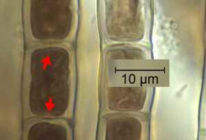
The evidence that demonstrates conclusively that this is a red alga can be seen in the image below. A gap in the cell walls often remains between daughter cells after cell division, which means that the protoplasm of the two cells is connected.   As the cells develop, these “pit connections” become filled by “pit plugs”, formed from protein and polysaccharide, which are just visible in the light microscope.   Not all red algae have pit plugs, but it is particularly characteristic of the class Floridophyceae, to which Audouinella belongs.

Audouinella_pygmaea_pits

A high magnification view of filaments of Audouinella pygmaea from Anghidi Fawr stream, May 2015, with arrows indicating the pit connections between adjacent cells.

A few months ago, I wrote about the problems of knowing whether algae were genuinely rare or simply not very well recorded (see “A “red list” of endangered British diatoms?”). The Freshwater Algae Flora of the British Isles refers only to a single record of Audouinella pygmaea (River Deveron, Scotland), whilst my old copy of West and Fritsch also mentions a record at “Penyghent” in West Yorkshire.   Audouinella pygmaea is a good example of an alga that is probably under-recorded.   These tiny dark-brown spots are very easily overlooked by all but the most observant surveyors.   I suspect that it is more common than we think but that it is overlooked by both macrophyte surveyors (who are generally focussed on larger organisms) and diatom samplers (whose methods preclude the study of soft-bodied algae). A map of the current distribution of Audouinella pygmaea in the UK will, therefore, give an indication of where eagle-eyed surveyors are work than reveal any deep biogeographical truths.

Postscript: this post has been extensively re-written as closer observation by Dave John led us to change our initial conclusions about the identity of the alga. As Chris Carter pithily put it as we mulled over the issue, “anyone who does not make mistakes does not make anything”

References

John, D.M., Whitton, B.A. & Brock, A.J. (2011). The Freshwater Algal Flora of the British Isles. 2nd Edition. Cambridge University Press, Cambridge.

West, G.S. & Fritsch, F.E. (1927). A Treatise on the British Freshwater Algae.   Cambridge University Press, Cambridge.